Logo of Sri Balaji Hospitals
Book Consultation

Department of Arthroscopy
Shoulder Arthroscopy

Gain enhanced mobility with arthroscopic shoulder surgery at Sri Balaji Hospital, Chennai. Trust our experts for superior care and recovery.
Book an Appointment
A patient holding his shoulders because of pain.

The Shoulder Joint

The shoulder joint is one of the two ball and socket joints in the body (the hip joint being the other. It is one of the most complex and flexible joints of the body, as it offers movement in almost every plane and every direction. However, it is this high degree of flexibility and allowance of movements that also makes it so susceptible to injuries & dislocations.

Anatomy of Shoulder Joint

Boney Anatomy
Soft Tissue Anatomy

The shoulder is made up of:

  • The Humeral head (the ball)
  • The Glenoid of the scapula (the socket)
  • The Clavicle (collar bone).
  • Capsule: It encloses the ball & socket joint. The capsule’s primary function is to provide stability to the joint. Overuse or injuries of the capsule can result in capsular tears.
  • Bursae: The bursae are fluid-filled sacs that help in cushioning the joint from friction between muscles, tendons and even bones. Overuse or tear of surrounding muscles can result in inflammation of the bursae and pain (bursitis). The commonest form of bursitis is observed in a condition known as Sub-acromial bursitis.
  • Labrum: It is a fibrous cartilage which is rigid and lines the border of the glenoid or “cup” therein, providing depth and enhancing the stability of the shoulder joint. A tear in the labrum can cause sports injury.
  • Ligaments: These are thick connective tissues which provide bone-to-bone connections to keep the various joints in place. Excessive stretching during sports or injuries such as 2-wheeler accidents to the shoulder can result in ligament strain or ligament tears.
  • Muscles and tendons: These aid in providing strength & mobility to the shoulder joint. Excessive weight lifting or impact injuries following road traffic accidents can result in the tearing of muscles and tendons. The most important of these muscles are a group of muscles known as the “rotator cuff”. Tear of the rotator cuff is one of the commonest indications for persistent pain without an actual injury (e.g., Age-related (degenerative) tears, tears due to overuse/ sports).

What is Shoulder Arthroscopy?

Shoulder arthroscopy is a surgical procedure in which an arthroscope is inserted into the shoulder joint to view the entirety of the joint and detect/ repair/ reconstruct injuries associated with the finer injuries/ diseases which do not recover the traditional “open surgery” technique.

Contact usA patient in pain caused by a shoulder problem.
When to Opt for Shoulder Arthroscopy
Shoulder Arthroscopy Procedure
Advantages of Shoulder Arthroscopy
Recovery after Shoulder Arthroscopy

NOT ALL shoulder problems/ injuries require surgery or even a keyhole surgery. In fact, most arthroscopic or keyhole surgeries are suggested only when all forms of non-operative treatment have failed or are forecasted to be non-beneficial for the individual.

Arthroscopic or keyhole surgery is often the preferred surgical approach for the following shoulder problems, including but not limited to:

  • Diagnostic arthroscopy: Evaluation and treatment of instability.
  • Debridement/drainage of shoulder joint infection
  • Biceps tendon tear, which will not heal via non-surgical means.
  • A torn rotator cuff
  • Stiffness of the shoulder
  • Subacromial decompression
  • Arthritis of the end of the clavicle
  • Frozen shoulder or adhesive capsulitis.
  • Removal of loose bodies
  • A torn or damaged ring of cartilage (labrum) surrounding the socket lining the area where the head of the humerus (ball of the joint) is seated.

An arthroscope is a small fiber-optic device which comprises several parts, such as:

  • Tiny lens
  • A light source
  • A mini 4K definition video camera linked to a 4K monitor.

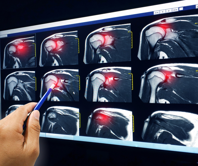
Here at Sri Balaji Hospital, we use the state-of-the-art Smith & Nephew arthroscopic system with 4K quality to visualise the affected joint as a whole. It can screen for associated injuries (sometimes missed even during MRI studies).

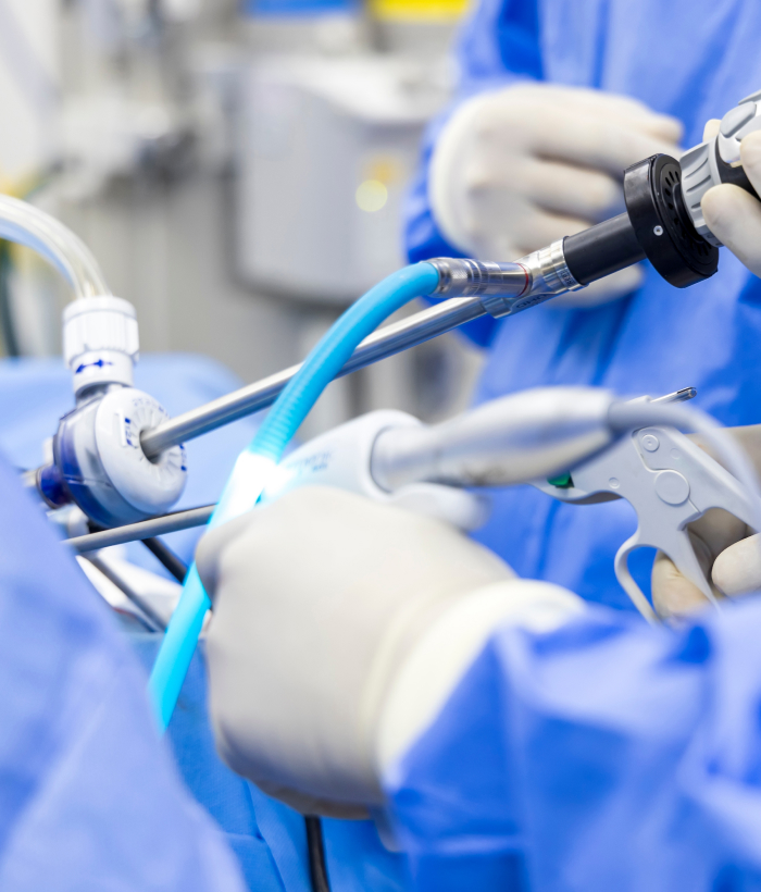
Small incisions (known as portals) are made around the joint area. Through one portal, the arthroscope (the viewing device) is inserted to view the shoulder joint. The other portal is used for the insertion of surgical instruments to probe various parts within the joint and repair the damaged shoulder.

Arthroscopy is a far less invasive method of exploring the muscles, ligaments and tissues and the damage to them. Also, in contrast to open surgeries, the approach and surgery itself cause little to no damage to the surrounding non-damaged areas when compared to traditional open surgeries. However, it must still be noted that arthroscopic surgeries have specific indications for use, as mentioned above.

Here at Sri Balaji Hospital, we follow specifically designed protocols for post-operative rehabilitation, taking into account the

  1. Age of the patient
  2. Type of injury to the shoulder joint.
  3. Extent of injury/ disease of the shoulder joint.

Conditions not involving tears, such as sub-acromion decompression, arthroscopic release of frozen shoulder, arthroscopic removal of loose bodies, or debridement, allow individuals to resume shoulder mobilisation within the first 24 hours following the surgery.

For arthroscopic repairs of torn elements (rotator cuff, labral tears, recurrent dislocation repair surgeries) of the shoulder, rehabilitation involves the following protocol as devised by our expert team at Sri Balaji Hospital:

Within 24 hours: Discharge to home

Up to 2 weeks: Ice packs and gentle passive movement of the operated shoulder with limitless movement to the elbow and wrist of the same side. Patients can start having a bath immediately from day 1 of surgery.

At 2 weeks: Suture (stitch) removal with passive movement of the operated shoulder up to 45 degrees.

2 to 4 weeks: Active range of movements up to 45 degrees and passive movements between 45 & 90 degrees are encouraged.

4th to 6th week: Unrestricted movement in all planes and directions.

Images showing rotator cuff tear.

Rotator Cuff Tears

Rotator cuff muscles are a group of muscles entrusted with the function of lifting and rotating the arm. The rotator cuff is made up of the following muscles:

  • Supraspinatus muscle.
  • Subscapularis
  • Infraspinatus
  • Teres minor

Although the rotator cuff is a strong muscle group, cuff tears can occur as a result of thinning of the muscles with progressive age (degenerative cuff tears) or due to injury.

Classification of rotator cuff tears

The severity of Rotator cuff tears is graded based on the size of the tears. They are

  1. Small or partial tears
  2. Large tears
  3. Massive tears.
Do all Rotator cuff tears need surgery?
NO. Small or partial tears often do very well without surgery. However, larger tears will benefit from arthroscopic surgery, while massive tears may even require open surgery. The need for surgery (or not), like most other arthroscopic surgeries, is based on the patient's age and the demands placed on the shoulders prior to the injury.
What is the typical timeframe for returning to sports and heavy lifting after a cuff repair?
We at Sri Balaji Hospital believe in early mobilisation to reduce the risk of long-term stiffness. Therefore, we initiate movement of the operated shoulder anytime between 24 hours post-surgery to 14 days post-surgery (depending on the extent of the tear). Rehabilitation will progressively be stepped up every two weeks, and we expect a full return to sporting activities and weight training in 3 to 6 months at the most.
Aim Of Surgery
Post-Surgery Rehabilitation Protocol
Arthroscopic surgery aims to repair the cuff tear with high-quality medical grade material via arthroscopic (keyhole) surgery to hold the torn ends of the cuff together to aid resisting forces until healing of the torn rotator cuff is achieved. The materials used in cuff repairs are usually “dissolving” or bio-absorbable screws with non-dissolving (non-absorbable) suture anchors. Following the initiation of the healing process, physical therapy and rehabilitation are stepped up on a “2-weekly basis” to restore flexibility and range of movement. At the same time, the operated cuff remains intact and continues to heal.
Within 24 hours
Discharge to home
Up to 2 weeks
Ice packs and gentle passive movement of the operated shoulder with limitless movement to the elbow and wrist of the same side. Patients can start having a bath immediately from day 1 of surgery.
At 2 weeks
Suture (stitch) removal with passive movement of the operated shoulder up to 45 degrees.
2 to 4 weeks
Active range of movements up to 45 degrees and passive movements between 45 & 90 degrees are encouraged.
4th to 6th week
Unrestricted movement in all planes and directions.
Medical experts performing Arthroscopic Surgery.

Arthroscopic Surgery for Shoulder Instability / Recurrent Shoulder Dislocations

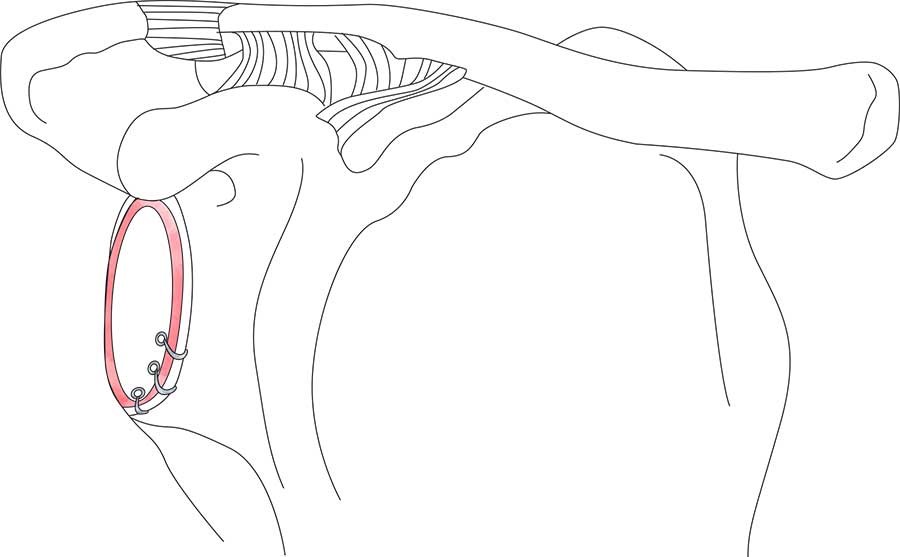
Most patients who suffer a dislocation of the shoulder following an injury often suffer a tear of the labrum (which lines the socket of the shoulder to provide depth & thereby provide stability).

These tears (known as labral tears) can affect the stability of the head of the humerus (the ball which lies within the socket), resulting in instability and recurrent dislocations.

Patients with recurrent shoulder dislocations may have an isolated labral tear or associated injuries to the ball & socket joint, such as chipping off of the bone of the socket or flattening of the ball (due to frequent sliding out of the ball through the surface of the socket) which further increases the “wear & tear” and also the risk of recurrent dislocations.

The arthroscopic Bankart repair is a procedure which helps in repairing this labral tear, thus bringing back the stability to the shoulder joint. In isolated labral tears, this surgery alone is sufficient to bring back the desired stability.

However, when there is a combination of injuries, such as the chipping of bone or flattening, an additional procedure, known as remplissage, may be required. In case of large bony defects, an open procedure known as the latarjet may be necessary to bring about all-around stability to the shoulder joint.

Procedure
What to Expect After Surgery

The arthroscopic Bankart repair is performed using advanced bio-composite bone anchors that are fastened into the Glenoid (also known as the socket). The site of these anchors allows their eventual replacement by bone. The bone anchors have non-dissolving sutures attached to them. These sutures are then used to tie the torn labrum back to where it has torn off from the front of the socket.

The diagrammatic representation shows how a labral tear looks before and after Bankart's (labral) repair.
The above diagrammatic representation shows how a labral tear looks before and after Bankart (labral) repair.
Within 24 hours
Discharge to home
Up to 2 weeks
Ice packs and gentle passive movement of the operated shoulder with limitless movement to the elbow and wrist of the same side. Patients can start having a bath immediately from day 1 of surgery.
At 2 weeks
Suture (stitch) removal with passive movement of the operated shoulder up to 45 degrees.
2 to 4 weeks
Active range of movements up to 45 degrees and passive movements between 45 & 90 degrees are encouraged.
4th to 6th week
Movement in all planes and directions unrestricted.
Want to book an appointment with our doctors or know more about our medical facilities?
Contact Us
Logo of Sri Balaji Hospitals
Sri Balaji Hospital is a centre for Orthopaedics and Traumatology and is unwavering in its commitment to providing high-quality healthcare at affordable rates. Our dedication stems from the belief that you, the community, are at the core of every endeavour, deserving nothing less than the utmost care.
GET IN TOUCH
© 2024 All Rights Reserved. Powered by DigitalSEO
magnifiercrosschevron-downarrow-right